
Seminar Line Up
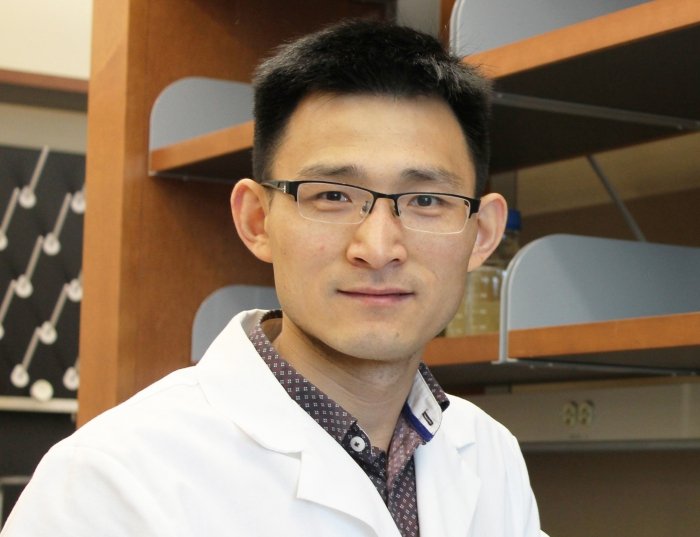
Anurag Paranjape, PhD
Dr. Paranjape is a Research Instructor in the University of Pittsburgh Heart and Vascular Institute and Vascular Medicine Institute.
Dr Paranjape’s research interest broadly focuses on identifying new ways to enhance drug-delivery in cardiovascular, neurological, and metastatic diseases. During his PhD training, he worked on identifying and characterizing unique, hard to target breast cancer stem cells. He generated in vitro transformed cell lines useful for studying epithelial-mesenchymal transition (EMT) and stemness properties, some of the hallmarks of cancer stem cells (Paranjape et al., Oncogene, 2012). Using these cells, he reported a novel mechanism linking Bmi1, Nanog, and NFκB in breast cancer stem cells (Paranjape et al., BMC Cancer, 2014). At MD Anderson Center, he demonstrated that FOXC2, a known EMT regulator, is involved in stem cell regulation in human prostate cancer cells (Paranjape et al., Oncogene, 2016). He showed that inhibition of p38 MAPK pathway using a chemical inhibitor reversed the EMT and made the cells more sensitive to AR therapy. Later at NCI, he worked on ‘hard to treat’ breast cancer brain metastases for which no effective therapy exists. That study showed that distinct areas within breast cancer brain metastases with varying permeability for drugs have unique gene expression patterns and identified that astrocytic S1P3 is upregulated as a neuroinflammatory response in highly permeable lesions (Gril and Paranjape et al., Nature communications, 2018).
Currently he is working on establishing technologies to increase drug delivery beyond the endothelial barriers in the heart and brain which have been known as hindrances in treating cardiovascular and neurological diseases. His is exploring the probable bioeffects of new non-invasive technologies such as ultrasound-targeted microbubble cavitation (UTMC) to induce hyperpermeability.

Rodrigo Machado-Florentino, PhD
Liver-Related Work
Recently some point mutations have been associated with specific liver diseases. My interested is to understand the relationship between these mutations and the incidence of liver disease to find out the mechanism behind it. With the advances in induced pluripotent stem cell (iPS cell) technology and the ability to differentiate these cells into hepatic cells, such as hepatocytes, cholangiocytes and stellate cells, a new and a better model is available to study human liver diseases.
Yashika Kamte, PhD
Dr. Kamte is am a junior postdoctoral scholar in the laboratory of Dr. Michael Wells at the University of California, Los Angeles. She completed her PhD from Duquesne University in Pittsburgh under the mentorship of Dr. Lauren A. O'Donnell. Her current research focus is to identify various ways pathogens can disrupt neurodevelopment and lead to abnormalities. In the Wells lab her goal is to identify if genetic heterogeneity can determine the fate of neural stem cells in the face of environmental perturbations such as viral infections. Specifically, she wants to determine if specific single nucleotide polymorphism (SNPs) can make a donor more or less sensitive to the impacts of infection and inflammation. To do this, we employ novel platforms such as "cell villages" and "chimeroid villages" where we pool several donor hiPSC derived NPCs into a single cell culture environment and then use computational techniques to identify what cells/transcriptomics belong to a specific donor.
For her talk, she will focus on how we construct cell villages and chimeroids and then focus on how to use these models to answer their research questions.
For more information on the Wells lab and the research we do you can check out our lab website - https://wellslab.dgsom.ucla.edu.
Source: wellslab.dgsom.ucla.edu

Jonathan Grasman, PhD
Dr. Grasman is an Assistant Professor in the Department of Biomedical Engineering at the New Jersey Institute of Technology.
Our research objective is to better elucidate the mechanisms behind tissue innervation and soft tissue reconstruction. Specifically, we focus on fabricating tissue systems from a variety of biopolymers to generate neurovascular and skeletal muscle tissue mimetics to enhance regeneration and innervation in incidents of traumatic injury, neuropathy, or genetic disorders both in in vitro systems and in vivo.
Some of the projects we are currently pursuing are:
Neural Engineering and Neurovascular Interactions
A major thrust of the lab is to develop 3D tissue models to understand the mechanisms of tissue innervation and neural network formation. We have developed tissue mimetics to study neurovascular interactions and neural network formation as a result of the interplay between the signaling cascades of vascular and neural networks. The model has identified several growth factors essential for the cross-talk between vascular and neural tissues, and we are characterizing traumatic injury models within this system to understand mechanisms behind neural repair and network reformation.
Skeletal Muscle Tissue Engineering
The second major thrust of the lab is to develop strategies to understand and augment skeletal muscle regeneration in craniofacial and limb settings. There is a significant need to develop strategies to treat volumetric muscle loss (VML) injuries, which occur from traumatic incidents ranging from car crashes to combat wounds. Our approaches include the development of off-the-shelf biomaterials to augment skeletal muscle repair, and also to develop in vitro mimetic tissues to understand the development and repair mechanisms behind skeletal muscle tissue formation.
Biomaterial Customization
Biomaterial synthesis and characterization is critical for successful tissue engineered strategies. We focus on biopolymer (e.g. fibrin, collagen, and silk fibroin) scaffolds and work to tailor the structural, mechanical, and biochemical properties of these materials to enhance therapeutic outcomes. Current foci are in the fields of vascular, neural, and muscle tissue engineering.

Lee Fortunato, PhD
Dr. Fortunato is a Professor in the Department of Biological Sciences at the University of Idaho.
The Fortunato lab seeks to understand the mechanism behind the development of morbidity and mortality in infants congenitally infected with human cytomegalovirus (HCMV).
Exploring the development of CNS defects in infants congenitally infected with HCMV. Studies in our lab focus on three important findings we have made regarding interactions with the host cell: 1) HCMV can inflict site-specific chromosomal damage to the host DNA; 2) HCMV dysregulates DNA repair in infected cells and 3) infection of neural progenitor cells by HCMV causes premature and abnormal differentiation of these important CNS cells. We hope to connect our observations in tissue culture to what occurs during development of the CNS in infected infants.
Interactions of HCMV with the cell cycle regulatory protein p53. Studies in the lab have shown that: 1) HCMV interacts with p53 and sequesters it within the viral replication centers in the infected cell nucleus; 2) in those centers, p53 binds site-specifically to the viral genome and 3) infection of p53 knockout cells show dramatic decreases in viral titers, in viral DNA replication and in viral protein expression. Future work will continue to characterize the many areas where p53 plays a role in the HCMV life cycle.


Kathy H.Y. Shair, PhD
Dr. Shair is an Associate Professor in the Department of Microbiology and Molecular Genetics at the University of Pittsburgh School of Medicine.
The Shair lab studies the Epstein-Barr virus (EBV) infection and EBV oncogenic mechanisms. EBV is a ubiquitous pathogen that is classified as a human tumor virus because it is associated with the neoplastic cells of a number of B-cell lymphomas and epithelial cell carcinomas. Our goal is to understand the host and viral molecular determinants of EBV oncogenesis in human cells and experimental culture models of EBV infection. A major focus of the lab is to understand the molecular pathogenesis of EBV in nasopharyngeal carcinoma. There are three primary research projects: 1) elucidating the mechanism by which EBV oncogenic proteins modulate the DNA damage response, 2) developing experimental organoid models of the nasopharyngeal epithelia to understand early infection events in the nasopharynx, 3) identifying EBV serological biomarkers for risk assessment and the early detection of EBV-associated cancers. There is currently no FDA-approved prophylactic vaccine that can prevent EBV infection. Given that EBV infection is persistent for the lifetime of the host and the high seroprevalence in the adult population, our ultimate goal is to understand why some individuals could be at risk of developing EBV-associated cancers.

Susana da Silva, PhD and Jincy Joy, PhD
Dr. Susana da Silva is an Assistant Professor of Ophthalmology and Developmental Biology at the University of Pittsburgh School of Medicine.
The research in Dr. da Silva’s laboratory is centered on a small highly specialized area of the retina named the fovea. The fovea is our high acuity area and is responsible for our ability to perform tasks such as reading, driving and recognizing faces. This area is very distinct from the rest of the retina presenting unique and specialized cellular and functional properties. Trained as a neurodevelopmental biologist, Dr. da Silva is very interested in deciphering the molecular developmental mechanisms orchestrating the formation of such specialization in the retina. To achieve this Dr. da Silva’s laboratory uses a multidisciplinary research program based in multiple model organisms, combining state-of-the-art genomic, molecular, and tissue culture techniques. The overarching goal of Dr. da Silva’s research is to advance the current understanding of basic genetic and molecular mechanisms underlying fovea development and subsequently establish new experimental models of human foveal diseases with wide applicability and therapeutic potential.
Dr. Jincy Joy is a Postdoctoral Associate in the Department of Bioengineering, Swanson School of Engineering with Prof. Jonathan Vande Geest at the University of Pittsburgh School of Medicine.
Her background in chemistry and training as a biomedical engineering during her PhD at the Indian Institute of Technology Delhi, India aligned her research interest in designing natural polymer-based bioresorbable biomaterials for tissue engineering and regenerative medicine. Understanding the importance to control the interaction of biomaterials with the innate immune system, she designed liposomes for targeted delivery to macrophages as a postdoctoral fellow at the Max Planck Institute for Medical Research, Germany.
Her expertise as a biomaterial scientist brought forth this scientific collaboration with the da Silva lab in designing a novel biocompatible gelatin-based Bruch’s membrane-retinal organoid assembloid. This assembloid is designed to serve as an in-vitro platform for studying the mechanistic cellular interaction of human induced-pluripotent stem cell reprogrammed retinal pigment epithelium (hiRPE) cells, and photoreceptors which plays a pivotal role in vision loss in age-related macular degeneration (AMD). This study will enable tailoring a patient-specific assembloid to develop personalized medicine to impede vision loss in AMD.

Daniel Shiwarski, PhD
Dr. Shiwarski is an Assistant Professor within the Department of Bioengineering and the Vascular Medicine Institute at the University of Pittsburgh. He obtained a combined undergraduate degree from Bucknell University in cell biology and biochemistry, a Ph.D. in Biological Sciences and Neuroscience from Carnegie Mellon University and the Center for Neural Basis of Cognition, and completed a Postdoctoral fellowship in Biomedical Engineering at Carnegie Mellon University in cellular biomechanics and tissue engineering.
The key innovation in Freeform Reversible Embedding of Suspended Hydrogels (FRESH) 3D bioprinting is the extrusion and embedding of an ECM hydrogel within a support bath that holds the extruded gel in place during printing.
In a recent publication in Science we showcased FRESH by 3D printing collagen-I into functional components of the human heart. Multiple bioinks can be printed including alginate, fibrin, collagen-I, methacrylated hyaluronic acid, decellularized ECM, and Matrigel demonstrating our ability to utilize a range of bioinks to tune scaffold composition.
Recently, we created a collagen-based 3D bioprinted vascular microfluidic platform using FRESH bioprinting (CHIPS) as well as the development of a custom perfusion bioreactor for long-term perfusion culture (VAPOR). Together, these systems establish the foundation for design and fabrication to accurately and reproducibly bioprint any 3D fluidic channel design within a collagen-scaffold. At the University of Pittsburgh in the Departments of Bioengineering and Vascular Medicine Institute we collaborate with many investigators from the School of Medicine and Carnegie Mellon University to incorporate physiological readouts, bioelectronics, and real-time fluorescence imaging into our platforms for organ-on-chip systems to study human vascular, kidney, and pancreatic disease.

Afshin Beheshti, PhD
Dr. Afshin Beheshti is a Professor at the University of Pittsburgh School of Medicine, Director of the Space Biomedicine Program at University of Pittsburgh, and the Associate Director at the McGowan Institute of Regenerative Medicine at University of Pittsburgh.
Dr. Beheshti earned his PhD in physics from Florida State University in 2002 before transitioning to postdoctoral training in cancer research, systems biology, space biology, and radiation biology. From 2006 to 2011, he was part of a NASA Specialized Center of Research (NSCOR) funded project, focusing on cancer risks due to space irradiation, during which he published several key studies. During this time, he also explored how aging affects tumor growth dynamics using novel systems biology approaches, alongside other cancer-related research. In 2014, Dr. Beheshti joined Tufts University School of Medicine/Tufts Medical Center as an Assistant Professor, where his research as a systems biologist centered on cancer, particularly in areas such as microRNAs (miRNAs), aging, cancer drug targets, and the development of novel immunotherapies. In April 2017, he joined KBR at NASA Ames Research Center, contributing to the GeneLab project and platform development. Shortly thereafter, he secured multiple grants from NASA, the Department of Defense, and other agencies, leading research on the roles of miRNAs and mitochondria in space biology, countermeasures against space radiation and microgravity, COVID-19, Long COVID, cancer, and the effects of high altitude on human biology. In 2023, Dr. Beheshti transitioned to the Blue Marble Space Institute of Science at NASA Ames Research Center, where he continues to advance research in these critical areas.
He recently joined the University of Pittsburgh as a Professor at the School of Medicine, Director of the Space Biomedicine Program, and Associate Director at the McGowan Institute of Regenerative Medicine. In these roles, he will continue his ongoing projects and launch a new space biomedicine program at the University. With this new initiative he will create research opportunities to explore space health issues, research countermeasures to mitigate the damage caused by the space environment and develop outreach/education programs for space biology research. Additionally, he holds a Visiting Researcher appointment at the Broad Institute of MIT and Harvard. Dr. Beheshti is also the President of two non-profit organizations. The first, the COVID-19 International Research Team (COV-IRT, www.cov-irt.org), was formed in March 2020 and focuses on COVID-19 and Long COVID research, with many publications produced. The second, established in August 2023, aims to democratize access to artificial intelligence through the design, construction, and maintenance of a free Personal AI called Kwaai (www.kwaai.ai).
Recently, Dr. Beheshti, was one of the leads who partnered with Nature Portfolio to coordinate the largest-ever collection of space biology papers, the Space Omics and Medical Atlas (SOMA). This project involved over 100 institutions from more than 25 countries, resulting in an extensive compendium of manuscripts, data, protocols, and code for aerospace medicine and space biology. Dr. Beheshti contributed to 22 papers in this collection, serving as senior author on nine. His contributions were pivotal in securing the cover feature for the August 2024 issue of Nature, which included three of the papers he co-authored, with one highlighting his role as senior author. The issue can be found here: Nature August 2024.
Dr. Beheshti's contributions have earned him recognition and accolades, including the International Space Station Research & Development Award for Compelling Results in Biology from the American Astronaut Society/NASA, the NASA Exceptional Scientific Achievement Medal, One KBR Award, and the NASA Outstanding Service Award: NASA Ames Safety Award Program II.
Polly Mattila, PhD, and Adrian Lee, PhD
A special seminar focused on the new offerings of the University of Pittsburgh Stem Cell Core-arriving summer 2024.

Sam Moss
Samuel Moss is a fourth year PhD candidate in Dr. Adam Feinberg’s Regenerative Biomaterials and Therapeutics Group. He earned his BS in Biomedical Engineering from the University of Wisconsin – Madison in 2020. His work is primarily focused on fabricating structured organoids and cell aggregates using FRESH 3D bioprinting.
Ioannis K. Zervantonakis, PhD: Microfluidic approaches to study cancer-macrophage interactions in 3D environments
Dr. Ioannis Zervantonakis is an Assistant Professor in the University of Pittsburgh Bioengineering Department, and a member of the UPMC Hillman Cancer Center. Prior to this, Dr. Zervantonakis was at Harvard Medical School where he was a Postdoctoral Research Fellow and Instructor in Cell Biology.
Dr. Zervantonakis runs the Tumor Microenvironment Engineering Laboratory (TME Lab) where they employ a quantitative approach that integrates microfluidics, systems biology modeling, and in vivo experiments to investigate the role of the tumor microenvironment on breast and ovarian cancer growth, metastasis, and drug resistance. The goal of the TME lab research program is to discover biomarkers that guide new drug development and improve prognosis; develop new strategies to optimize existing treatment protocols, and engineer microfabricated tools that enable screening and personalization of cancer therapies. Projects in the TME Lab include: 1.) microfluidic and microfabricated assays to model the tumor microenvironment and study tumor, fibroblast and immune cell infiltration; 2.) systems biology approaches to cancer drug resistance and metastasis in breast and ovarian cancer; tumor heterogeneity: 3.) single cell phenotypic decisions; and 4.) localized drug release and gradients within the tumor microenvironment

Liz Johnston, PhD Candidate (Abbot Lab, CMU)
Liz Johnston is a Ph.D. student in Dr. Rosalyn Abbott’s Lab within the Department of Biomedical Engineering at Carnegie Mellon University. Her work focuses on the development of an efficacious defatting cocktail to defat steatotic livers prior to liver transplant. In her talk, she will discuss the production of a three-dimensional model of steatosis that mimics the pathology seen in discarded steatotic livers. She will then show her approach toward the development of an effective defatting cocktail for future incorporation into normothermic machine perfusion systems to improve steatotic liver transplant outcomes.
The Abbott Lab is in the Departments of Biomedical Engineering and Materials Science and Engineering at Carnegie Mellon University.

Megan Culler Freeman, MD, PhD: Spinal Cord Organoids as a Model for EV-D68 CNS Infection
Dr. Megan Culler Freeman is an Assistant Professor in the University of Pittsburgh Department of Pediatrics and physician-scientist in the Division of Infectious Diseases.
The Freeman lab studies picornaviruses with tropism for the central nervous system using human tissue models (organoids) of relevant sites derived from induced, pluripotent stem cells.
Their current focus is on how enterovirus D68 (EV-D68) mediates acute flaccid myelitis (AFM), a polio-like illness, in children by using a novel human spinal cord organoid model.
AFM results in moderate to severe paralysis in previously healthy children due to spinal cord damage, leading to lifelong increased medical needs. It is unknown how EV-D68 causes paralysis in children and effective medicines for this condition do not exist. Understanding how EV-D68 infection leads to spinal cord damage could lead to new medications, prevention, or treatment strategies for these viruses, leading to improved care of children with AFM. Their work is also supported by patient-derived samples (human cell and viral) to better understand how specific viral genotypes and specific host factors contribute to pathogenesis of this rare condition.
Guang Li, PhD: Study of heart development and congenital heart defects using iPSCs and organoids
Guang Li earned his PhD in genetics from the Chinese Academy of Sciences, where he worked on the epigenetic regulation of plant phase development. He then completed postdoctoral training at Stanford University, studying heart development using single-cell approaches. Dr. Li generated the first single-cell RNA-seq atlas of the early mouse embryonic heart. He joined the faculty at the University of Pittsburgh in 2019 and has co-authored 29 peer-reviewed publications. Dr. Li was also awarded the NIH Director's New Innovator Award. Currently, his lab focuses on single-cell analysis of developing mouse hearts and creating four-chambered organoids using human iPSCs. Additionally, his lab models congenital heart defects such as Ebstein's anomaly and studies neonatal heart regeneration.
Dr. Li’s lab is in the Department of Developmental Biology at the University of Pittsburgh.
Velpandi Ayyavoo, PhD: Brain Organoids- An Emerging Model System to Study HIV-1 Neuropathogenesis.
Studying neurodegenerative diseases is complex and quite fascinating. Understanding how HIV-1 affects the brain and results in the development of HAND is the major focus of our laboratory. However, it is rather difficult due to the inability to obtain primary tissues or cells (neurons, astrocytes) from patients. One of the approaches is the use of 3D organoids that is very relevant to the physiological conditions and cell lineages observed in the brain. In an effort to develop such an organoid model, we propose to use three cell types (microglia, neurons, and astrocytes) that are known to play a critical role in HIV CNS infection and pathology. These cells will be derived from neuronal progenitor cells (NPCs) by various differentiation methodologies that will mimic the adult brain (see figure below showing primary Neurons and Astrocytes developed from NPCs). Studies are in progress to develop 3D culture and to further understand how HIV-1 alters the function, viability, and synaptic communication.
Dr. Ayyavoo’s lab is in the School of Public Health at the University of Pittsburgh.

Leonardo D'Aiuto, PhD: Modeling Host-Pathogen Interaction Using Induced Pluripotent Stem Cells
Dr. D’Aiuto’s area of expertise is neurotropic viruses, focusing on novel approaches for the generation of three-dimensional neuronal cultures from human-induced pluripotent stem cells to as a model system for exploring the relationship between central nervous system neurotropic viral infections and psychiatric disorders. Through a recently awarded R01 grant funded by the National Institute of Neurological Disorders and Stroke, Dr. D’Aiuto studies the effects of herpes simplex virus infection on neural progenitor cell biology. In addition, he serves as a co-investigator on federally and privately funded research projects with colleagues in Psychiatry, as well as Medicine, and in Infectious Diseases and Microbiology at Pitt Public Health.
Neil Hukriede, PhD: Using Human Kidney Organoids to Model Renal Injury
Acute kidney injury (AKI) is associated with a high mortality and morbidity and AKI survivors often develop end stage renal disease. At present, there are no established therapies to prevent renal injury or accelerate the rate of renal recovery following AKI. The consequences of abnormal kidney function are frequently fatal, with dialysis and organ transplantation the only current long-term treatments for kidney disease. Importantly, the vertebrate kidney has the potential to regenerate, but the molecular mechanisms of kidney regeneration are largely unknown. A better understanding of the processes controlling renal regeneration after injury may provide important clues for the development of new therapies.
The Huckriede lab is in the Department of Developmental Biology at the University of Pittsburgh.
Kalil G Abdullah, MD, MSc, FAANS: Finding the Limit of Patient-Derived Glioma Organoid Modeling
The Abdullah laboratory at the University of Pittsburgh is focused on developing novel clinical models of glioma and identifying druggable targets to facilitate early-phase clinical trials. Gliomas are intensely heterogeneous tumors that not only contain numerous cell types but also demonstrate the ability to transition between different phenotypic states. This complexity has made developing model systems that recapitulate human tumor biology both difficult and essential. Traditionally, models of gliomas are two-dimensional cell lines and only represent certain subtypes of the highest-grade glioma, glioblastoma. This is because the unique biology of lower-grade gliomas has prevented them from being studied either outside of the lab or in animals. We have created ex-vivo culture systems that allow us to investigate critical aspects of the tumor microenvironment, immune response, and discover targets for therapy. Our laboratory has previously shown the ability to establish lower-grade glioma organoids in vitro, maintain those cultures for extended periods of time, hibernate, and then reanimate tumor tissue without loss of either genetic or phenotypic fidelity. Our work also includes extensive and sophisticated live-cell imaging analysis that allows for longitudinal, non-invasive assessment of organoid response to treatment. Our organoid model systems, in addition to glioma stem cell and mouse models, allow us to perform highly sophisticated assessments of drug response across platforms and identify rare but critical druggable targets in gliomas. These analyses include complex metabolic tracing and immune cell response assessment. Despite the fundamental principles of genomics, immunology, and cellular cancer biology that underlie our work, our group focuses on projects that have a high potential for immediate clinical translation.

Open Forum
Space for discussion of technical challenges and series goals for the remainder of the year.

Amanda Clark, PhD: An Integrative Liver Microphysiological Tissue System for Modeling Metastatic Disease
The Clark lab at the University of Pittsburgh focuses on determining the molecular and cellular regulators of metastatic dormancy and recurrence within the liver. The lab utilizes a novel all-human ex vivo 3D liver microphysiological system to model metastasis. The system has not only enabled the recreation of dormant-emergent metastatic cancer progression as observed in vivo but also the identification of mechanisms, candidate biomarkers, and new therapeutic opportunities to target the various stages of metastasis. Their current research centers on: i) investigating how dysregulated gut homeostasis drives emergence from metastatic dormancy in the liver, and ii) examining how the bi-directional crosstalk mediated by extracellular vesicles regulates metastatic breast cancer dormancy in the liver.
Charlie Ren, PhD: Engineering Lung Tissue Morphogenesis
Dr. Ren was trained as a developmental biologist studying vertebrate embryogenesis. Moving from developmental biology to bioengineering, over the past ten years, he combines extracellular matrix and stem cell engineering to regenerate functional, vascularized tissues (lung, intestine and islet) to deliver specific biological functions. Current research in his lab at CMU investigates and engineers cell-cell and cell-extracellular interactions during the biofabrication of functional tissues, in particular the lung. Their recent work describes strategies for modeling human heart and lung co-development using stem cell derived multi-lineage differentiation and for precise control of epithelial polarity in engineered lung organoid. These topics will be covered in this talk.

Adrian Lee, PhD: A Living Biobank of Breast Cancer
The Lee/Oesterreich lab at the University of Pittsburgh studies the molecular basis of breast cancer development and resistance to therapy, with the goal to improve precision medicine and outcomes for breast cancer patients. The laboratory employs a systems biology approach, utilizing a combination of single cell and bulk sequencing, computational methods and biological models to identify and validate new drivers and therapeutic targets. Hypotheses are tested in vitro and in vivo and then moved to clinical trials.
The majority of studies incorporate analysis of human specimens, in collaboration with a large network of clinicians and nurses. This includes computational analysis and modeling of large biomedical and genomic datasets including electronic health record data.
A major focus of the laboratory is identifying mechanisms of resistance to endocrine therapy, and new approaches to blocking breast cancer metastasis through precision medicine. This includes the study of estrogen receptor (ESR1) mutations and fusions and synergism with growth factor pathways. A special focus is on the understanding of invasive lobular cancer (ILC), the second most common but understudied histological subtype of breast cancer.

Keisuke Ishihara, PhD: Topological Control of Organoids
The Ishihara lab takes a synthetic approach to study how cells form tissues. “Synthetic” embodies the experimental creation of states of physical organization and gene expression that push a multicellular system to all possible extremes. The synthetic approach allows us to discover novel regulatory molecules, dormant genetic programs, and general physical principles. Our goal is to develop new genetic and chemical tools to engineer form and function of human brain organoids and cardiac organoids. In this seminar, I will cover my work as a postdoc that used mouse neuroepithelial organoids to analyze and tune 3D tissue morphogenesis with a small molecule.